From Stanford Research, Dr. Paul Wong and associates have developed a 3D printed replica of the heart that is specific to the patient in need. It is fascinating to see the detail possible with the 3D prints. The structures that hold valves and the chambers of the heart give a kind of accuracy invaluable to doctors. Surgeons can adjust tools and materials to fit individual needs thanks to the replicas. They carry the kind of specificity for doctors to examine in detail a patient’s heart before they begin operating.
With a 3D printer, the doctor can leave the printer with the model sent from the computer and arrive the next day ready to take a close look and prepare for surgery. It is a new advantage that has the medical field excited. Having an exact replica of the heart is more valuable than a model on a screen because it can be observed and manipulated in hand in 3D without imagination. As Dr. Wong states, it allows doctors to test new methods on the model heart and work to a better solution before the patient lies on the operating table. After only a few hours, a print is available.
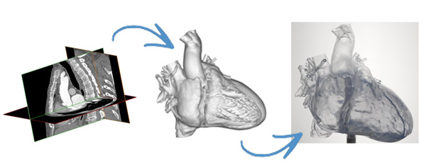
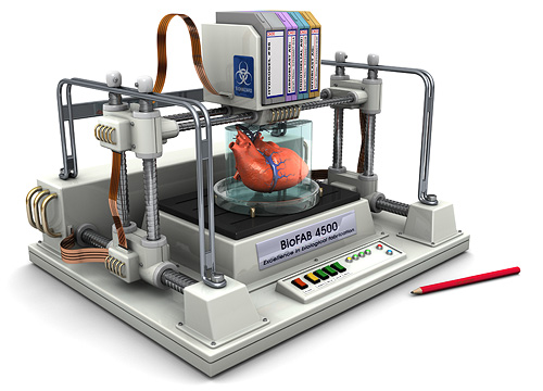
 As mentioned in the newscast, it is rumored that 3D printing is not far from printing actual organs. While such an achievement would be sensational, it is noteworthy to see the progress already available and see where 3D modelling for surgery can take future procedures and how doctors and patients can achieve the best possible outcome by having a clearer idea of options thanks to the 3D prints. After the scan, the images are downloaded into a software program translating the image into triangle format at a small enough grade allowing these fantastic replica hearts. It is one of the most complicated organs in the body, and 3D printing has it mapped and modelled. Before the chest is sawed open, it would be nice to see what will receive the surgeon’s touch, and now it is possible thanks to Stanford’s research team.
Source: ABC Local