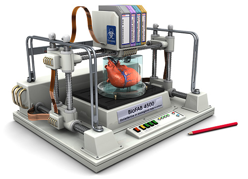
The risk, when writing about 3D bioprinting is that the “over-simplification” that is necessary to convey the news article’s message in the title gives an over-hyped idea of what has been achieved, with the inevitable let down that follows when we focus in on the actual facts. So, the recent video  Which brings us back to the process of developing the organoids, assembled by a 3D bioprinter using, in this case, an inkjet-like process. Dr. Atala’s team used genetically modified adult human skin cells and “transformed” them into pluripotent stem (IPS) cells. These can develop into any cell in the body and were “programmed” to become heart cell organoids. Each one has a diameter of about 0.25 millimeters and act as independent modules of a heart: when they receive the correct environmental cues, they beat.
In this specific case, the environmental cues were a special medium to keep them at the same temperature as the human body. The scientists also stimulated the miniature organ with electrical or chemical cues to alter the beating patterns. Growing them in three-dimensions through a 3D bioprinting systems allows them to interact with each other more easily, as they would in the human body. Although this project does not aim for the full reproduction of a human heart for implantation, it may help scientists find new ways to “assemble” human organs, without the use of a 3D printed polymer-based scaffold. Instead, they might find ways to even let complex organs self-assemble. In that case, we may have functional 3D printed organs even sooner than we think and the title of this article would, perhaps, turn out to to be an understatement in the end.

