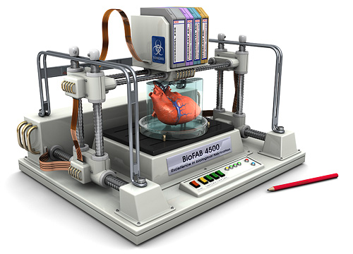
Credit: Organovo. “Organovo has described the first 3D bioprinted human kidney tissue, which is a significant advance over simple single layer cell cultures that are used today for drug testing and disease research. This image shows that the tissue is composed of different types of kidney cells that make up the proximal tubule, which plays a pivotal role in kidney disease. The tissue is 100% cellular (no scaffolds) with clear cellular connections and tissue architecture. (Not shown) The tissue also expresses normal kidney enzymes such as CYP450 (enzymes responsible for breaking down drugs) and gamma glutamyl transferase (GGT) activity, both of which are indicators of specific kidney cell (RPTEC) function. Key to colors and marks Tissues were stained with antibodies against two of the three different cell types found in the tissue: endothelial cell specific marker CD31 (green) and fibroblast marker TE7 (red). The third cell type is the polarized renal proximal tubular epithelial cells (RPTEC). The endothelial cells will associate with each other, forming networks. Over time, the center of these networks will “hollow out” to form microvessel lumens lined with epithelial cells. Networks with putative lumens lined with endothelial cells are marked (*).”
Organovo CTO and Executive Vice President of Research and Development, Dr. Sharon Presnell, explains, “Our bioprinted human kidney tissue represents a significant technical advance over the simple monolayer cell line cultures that predominate today. The histologic and functional features of the initial prototypes are compelling, and the in vitro durability of the system will likely enable the assessment of drug effects at chronic, physiologically relevant doses. Furthermore, the cellular complexity of the system will likely support mechanistic investigations into drug responses, including end points that have been difficult or impossible to assess in vitro, including tubular fibrosis and post-injury recovery.”
Speaking with Loyola University Medical Center’s Dr. Susan Hou, a well-regarded nephrologist who is also my mom, I was able to gain a better understanding of what this specific portion of the kidney is: “The kidney is made up of filtering units called glomerulae; they’re collections of capillaries that filter the blood, the function of the kidney. They filtered probably 100x what you need to get rid of. And there are tubules made up of four parts: the proximal, the loop of Henle, distal, and the collecting duct. These take back all of the waste that you don’t need to get rid of. So, it’s great to make proximal tubular cells, but, without the glomerulus, you can’t have any function of the kidney.”
She wasn’t able to comment specifically on the exhibited function of the epithelial cells demonstrated by Organovo’s research, as I was reading the company’s press release to her over the phone and probably mispronouncing “gamma glutamyl transferase”, but she did underscore the importance of the fact that they were able to print the “living interstitial layer comprised of renal fibroblasts (RF), and endothelial cells (EC)”, saying, “They’re a little bit closer to making the whole kidney if they’ve got the proximal tubule in the right position with the part of the kidney that’s between the filtering units.” In other words, Organovo is making significant progress, but still has some ways to go before 3D printing an entire, functioning kidney.